Discovery

Defects in a Cell’s Transport System Cause Rare Genetic Craniofacial Disorder

Defects in a Cell’s Transport System Cause Rare Genetic Craniofacial Disorder

The Finding

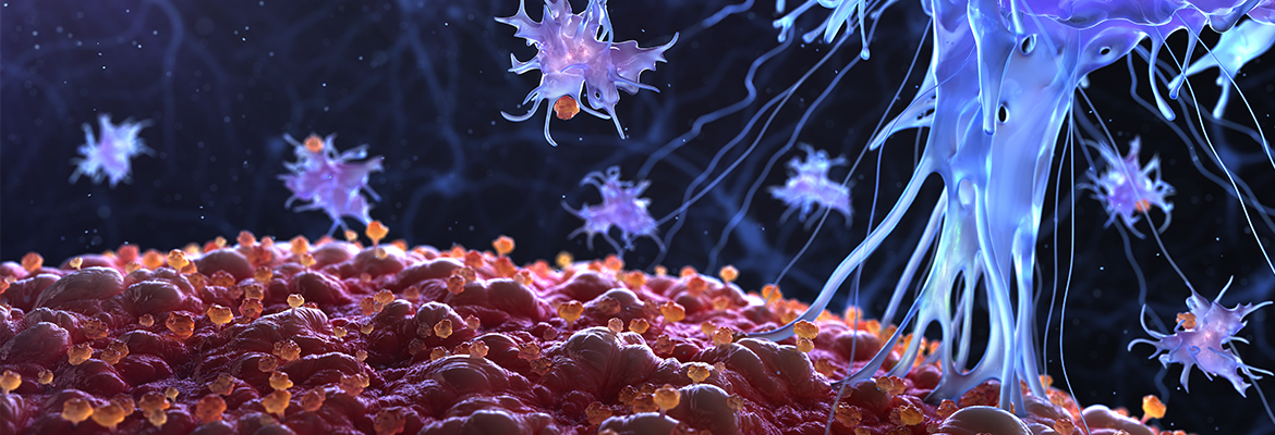
An international research team led by a Children’s Hospital of Philadelphia geneticist classified a new craniofacial disorder tied to disruptions in our cell’s tiny protein transport systems. The researchers identified the genetic disorder, ARCN1-related syndrome, in three children and an adult who share similarities in clinically recognizable facial differences, small heads and limbs, and mild developmental delays. As the condition’s name suggests, the four patients also share mutations in archain 1 (ARCN1) — a gene that encodes proteins in one of the body’s delivery systems for transporting, or “trafficking,” proteins between structures inside our cells: the COPI system. Previous research has linked the COPI system to neuronal functions, so the ARCN1 mutations may also affect brain development.

Why it matters

Families of children with rare disorders often experience stress as they embark on a series of tests and unsuccessful treatments known as “diagnostic odysseys” with no clear explanation for their complex condition. Putting a definitive name to a rare set of symptoms — and learning that other individuals share the same diagnosis — offers relief for patients while also bringing researchers one step closer to developing effective treatments. 

Who conducted the study

Kosuke Izumi, MD, PhD, attending physician in the Roberts Individualized Medical Genetics Center and the Division of Human Genetics at CHOP, led the study with colleagues from the University of Tokyo, Nagano Children’s Hospital, KK Women and Children’s Hospital, Shinshu University Graduate School of Medicine, Hopital Robert Debre, Centre Hospitalier Universitaire et Université de Liège, St Thomas’ Hospital, Genome Institute of Singapore, and SingHealth Duke-NUS Medical School.

How they did it

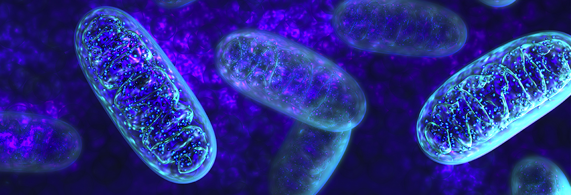
The researchers sequenced the exomes (the protein-coding portions of DNA) of the four patients and found loss-of-function mutations in the ARCN1 gene, which plays a role in the COPI system. COPI governs the transport of proteins between the cell’s endoplasmic reticulum (ER) and the Golgi apparatus so that when a mutation in ARCN1 disrupts the cell’s normal protein trafficking route, protein accumulates in the ER to cause a stress response that kills cells. ER stress can also impair the secretion of collagen — an important part of the connective tissue within bone and cartilage.

Quick thoughts

“This genetic syndrome represents a novel class of disorders,” Dr. Izumi said. “We have reported only the second known disease associated with the COPI protein complex, one of the three key intracellular protein transport systems.”

What’s next

Finding the cause of ACRN1-related syndrome is just the first step: The researchers will continue to identify more children and adults with similar symptoms, as well as conduct further studies to investigate COPI transport’s role in bone formation. “In order to provide better care for children with ARCN1-related syndrome, we still need to understand the full clinical spectrum of this disorder,” Dr. Izumi stated. “At the same time, we need to better understand the disease mechanism in order to discover therapies.”

Where the study was published

The study was published in the American Journal of Human Genetics.

Who helped fund the study

Grants from the Japanese governmental agency MEXT, Core Research for Evolutional Science and Technology, the Japan Agency for Medical Research and Development, the Singapore Ministry of Health, and the French Ministry of Health supported this research.

Where to learn more

You can learn more about this research in the press release and read the study abstract online. To find out about genetics research at CHOP, see the Roberts Individualized Medical Genetics Center and the division of Human Genetics.

Asthma and Allergic Rhinitis More Common in Children with Food Allergies

Asthma and Allergic Rhinitis More Common in Children with Food Allergies

The Finding

Children with a history of food allergies might also have a high risk of developing the most common childhood medical conditions in the U.S.: asthma and allergic rhinitis (also known as hay fever). Thirty-five percent of pediatric patients with an established diagnosis of food allergy went on to develop asthma, with the risk increasing for children with multiple food allergies compared to those with just one, according to new research published this year. Peanuts, milk, and eggs emerged as the most significant food allergens for predisposing children to asthma and rhinitis.

Why it matters

The disease rates of eczema, asthma, and rhinitis continue to fluctuate, with the current study finding higher asthma rates compared to previous reports. In Philadelphia, asthma affects one in five children— the highest rates in the nation. More surveillance and information about these conditions are necessary, and while previous research has already linked allergies to asthma, this is the largest study to date that examines the characteristics of primary healthcare provider-diagnosed eczema, asthma, allergic rhinitis, and food allergy in children.

Who conducted the study

The research team included David Hill, MD, PhD, fellow in the division of Allergy and Immunology, Robert Grundmeier, MD, attending physician and director of clinical informatics in the department of Biomedical and Health Informatics, Gita Ram, MD, attending physician in the division of Allergy, and Jonathan M. Spergel, MD, chief of the Allergy Section, all at CHOP.

How they did it

The researchers conducted a retrospective analysis of electronic health records from over one million urban and suburban children in the CHOP Care Network between 2001 and 2015. They divided the records into a closed-birth cohort of 29,662 children followed continuously for their first five years of life, and a cross-sectional cohort of 333,200 children and adolescents, followed for at least 12 months.

Quick thoughts

“Using provider-based diagnosis data provided important information often lacking in existing studies, ” said Dr. Spergel, senior author of the study in a press release. “We found different disease rates than previously reported, and our research provides key data to shape future efforts aimed at prevention, diagnosis and management of these common pediatric conditions. ”

What’s next

Further studies should investigate whether the food allergy patterns identified in their paper are comparable to data from other geographical areas including those found in rural areas (as the study cohort primarily consisted of children residing in urban or suburban settings).

Where the study was published

The study was published in BMC Pediatrics.

Who helped fund the study

The American Academy of Pediatrics Resident Research Award, the Stuart Starr Chair of Pediatrics, the Children’s Hospital of Philadelphia Food Allergy Research Fund, and Fare Education and Research Inc. supported the research.

Where to learn more

You can read the full study online at BMC Pediatrics or learn more in the press release. To learn more about allergy research at CHOP, see the Division of Allergy.

Gene Tied to Type 2 Diabetes May Inform Better Treatments

Gene Tied to Type 2 Diabetes May Inform Better Treatments

The Finding

Sophisticated scientific tools, including the gene editing tool CRISPR, allowed genomics researchers at Children’s Hospital of Philadelphia and the University of Pennsylvania’s Perelman School of Medicine to identify a new gene for Type 2 Diabetes (T2D) at a well-established genomic location. The gene in question, ACSL5, codes for a protein that regulates the body’s recognition of insulin — a crucial function that is disrupted in patients with T2D. Without the ability to properly use or make enough insulin, the body cannot move glucose into its cells. Developing drugs to act on the protein in question might help patients with T2D by increasing their sensitivity to insulin.

Why it matters

Type 2 Diabetes accounts for 90 to 95 percent of all diabetes cases, with an increase in cases for children and adolescents. Its exact cause, however, remains unknown. By learning more about the metabolic disorder’s biological mechanisms and complex factors — as well as environmental influences — researchers can develop more effective treatments. The study also illustrates the complexity of unraveling a disease through gene discovery, since the current study builds on past findings about T2D’s genetic underpinnings, including the role of another gene investigated in a previous study, TCF7L2.

Who conducted the study

The research team included investigators from the divisions of Human Genetics and Endocrinology, the division of Hematology, the department of Pathology and Laboratory Medicine, and NAPCore at CHOP, as well as the department of Pediatrics, the Institute of Diabetes, Obesity, and Metabolism, and the Institute for Biomedical Informatics at the University of Pennsylvania’s Perelman School of Medicine. Struan F.A. Grant, PhD, a genomics researcher at CHOP, led the study.

How they did it

Using CRISPR and a three-dimensional structural biology technique called circularized chromosome conformation capture (4C), Dr. Grant and his colleagues took a closer look at T2D from a molecular level — specifically, in cells derived from the colon. They used CRISPR to edit out precisely defined sequences around the TCF7L2 gene variant first reported by Dr. Grant in 2006 to be strongly associated with T2D in order to see how the deletion could change global gene expression. 4C also allowed them to examine the variant’s interactions with other gene locations. The researchers discovered that the TCF7L2 variant strongly influenced the expression of ACSL5, another gene.

What’s next

“This well-known genomic location harbors an especially strong signal, and may control multiple other genes, yet to be identified,” stated Dr. Grant in a press release. “In addition, we still don’t know which specific tissue or tissues that these T2D-related signals operate in to affect patients — whether they act primarily in the gut, in the liver, in adipose tissue or on beta cells in the pancreas. As we continue to better understand the biological mechanisms functioning in type 2 diabetes, we expect to find better strategies for treatment.”

Where the study was published

The study was published in Diabetologia.

Who helped fund the study

The Pennsylvania Department of Health, the Spatial and Functional Genomics Initiative at CHOP, and the Daniel B. Burke Endowed Chair for Diabetes Research supported this research.

Where to learn more

Learn more in the press release. To learn more about genetics research at CHOP, see the Division of Human Genetics.

Bio-Engineered Molecule Counteracts Effects of Newer Blood Thinners

Bio-Engineered Molecule Counteracts Effects of Newer Blood Thinners

The Finding

A novel bio-engineered molecule developed by hematology researchers at Children’s Hospital of Philadelphia and the University of Pennsylvania’s Perelman School of Medicine has the ability to quickly control bleeding in animal models when infused. If the factor, called FXaI16L, also proves to be effective in humans, it gives physicians a rapid countermeasure for child and adult patients who bleed extensively as a result of taking newer types of blood-thinning drugs. These blood-thinners, known as anticoagulants, are taken to counteract blood clots.

Why it matters

Blood clots play a role in heart attacks, strokes, and other serious conditions, killing one person every five minutes in the U.S. While anticoagulants can prevent these clots, they also increase the risk of bleeding. Thus, if a patient undergoes emergency surgery or suffers from a head bleed, doctors may need to reverse the anticoagulant’s effect with a countermeasure. The problem, however, is that the newer variety of blood-thinners, which are also deemed safer and more convenient than older ones like warfarin, currently do not have any approved countermeasures. In this study of injured mice modeled to have excessive bleeding, FXaI16L was able to rapidly control bleeding.

Who conducted the study

The study team involved researchers in the Division of Hematology, the Department of Pediatrics, and the Raymond G. Perelman Center for Cellular and Molecular Therapeutics at CHOP, and the department of Biostatistics and Epidemiology at the University of Pennsylvania’s Perelman School of Medicine. Rodney Camire, PhD, a hematology researcher in the Raymond G. Perelman Center for Cellular and Molecular Therapeutics at CHOP, led the study.

How they did it

Newer blood thinners work by blocking a key molecule in blood clotting, called factor Xa (FXa). With this in mind, Dr. Camire and his team of researchers developed the novel molecule, FXaI16L, by modifying FXa into a variant that is more potent, longer-lasting, and safer than naturally occurring FXa. To test the variant’s effectiveness, the researchers inhibited the expression of FXa in mice. When infused, the variant safely restored the mice’s blood clotting ability and significantly reduced bleeding.

Quick thoughts

“This molecule holds the potential to fill an important unmet clinical need,” stated Dr. Camire in a press release. “There are limited treatment options to stop uncontrolled bleeding in patients who are using the newer anticoagulant medications.”

What’s next

The next step involves testing the approach in large animals to determine whether the variant is indeed safe and effective enough to progress to a clinical trial, according to Dr. Camire.

Where the study was published

This study appeared in Nature Medicine.

Who helped fund the study

Researchers received support from the National Institutes of Health, the Penn-CHOP Blood Center for Patient Care and Discovery, and Pfizer Inc. Dr. Camire and his colleagues have been collaborating with Pfizer to develop the FXa variant to control bleeding for specific conditions, like intracranial hemorrhage. The molecule is currently in phase I clinical trials and is being conducted by Pfizer.

Where to learn more

You can read the study abstract online at Nature Medicine and learn more in the press release. To learn more about hematology research and care at CHOP, see the Division of Hematology.

Researchers Identify Gene Region That Raises the Risk for Ear Infections

Researchers Identify Gene Region That Raises the Risk for Ear Infections

The Finding

Middle ear infections, also known as acute otitis media (AOM), might be caused by microbes, but genetics can also make a child more susceptible to developing the painful condition. In the largest genomics study to date, researchers at CHOP and Rotterdam’s University Medical Center have discovered that a specific region in the human chromosome containing the gene FNDC1 raises a child’s risk for a middle ear infection.   

Why it matters

Ear infections are among the most common childhood illnesses, causing discomfort and stress for both young children and their parents. Discovering more about the condition’s biological mechanisms – including the interactions between genes and pathogens and who is most susceptible – gives researchers and clinicians a clue for developing more effective treatments. These treatments could include early medical interventions.

Who conducted the study

The study was led by Hakon Hakonarson, MD, PhD, director of the Center for Applied Genomics (CAG) with Jin Li, PhD, statistician at CAG as the lead analyst, along with collaborators led by Gijs van Ingen from the University Medical Center Rotterdam.

How they did it

The researchers conducted a genome-wide association study (GWAS) on two cohorts with DNA samples from 11,000 children. After discovering an association between middle ear infections and a site on chromosome six that contains the gene FNDC1, they replicated the finding in a separate cohort with data from 2,000 additional children. Further studies showed that in mice, the gene that corresponds to FNDC1 was expressed in the middle ear.

Quick thoughts

“Parents and pediatricians are all too familiar with this painful childhood ear infection — it’s the most frequent reason children receive antibiotics,” stated Dr. Hakonarson in a press release. “Although microbes cause this condition, it’s been well known that genetics also plays a role. This is the first and largest genetic study focused on risk susceptibility for acute otitis media.”

What’s next

In their paper, the researchers noted that learning more about the complex polygenetic pathogenesis of middle ear infections will, in the future, help to develop more specific therapies.

Where the study was published

The study was published in Nature Communications.

Who helped fund the study

The National Institutes of Health, the EU 7th Framework Programme, and the Kubert Estate family supported this research.

Where to learn more

Learn more in the press release and read the study’s abstract online. To learn more about CHOP research into the genetic causes of rare and complex childhood diseases, see the Center for Applied Genomics.

Pediatric Hypertension Often Goes Undiagnosed and Untreated

Pediatric Hypertension Often Goes Undiagnosed and Untreated

The Finding

This is the first study to show a widespread underdiagnosis of hypertension (high blood pressure) and prehypertension by pediatricians in U.S. children ages 3 to 18. The researchers found that only 23 percent of those who had blood pressures consistent with hypertension at multiple primary care visits were diagnosed with the disease, and only 10 percent of patients with symptoms of prehypertension were diagnosed. Of those children and adolescents with diagnoses of hypertension for at least a year, only 6 percent of those who needed anti-hypertension medication received a prescription.

Why it matters

Hypertension is one of the 10 most common chronic diseases in childhood, but it is often overlooked because symptoms are usually silent. High blood pressure levels can carry over into adulthood, and if left untreated, can lead to cardiovascular disease and damage to the kidneys and brain. Catching the disease early can help families introduce strategies such as lifestyle changes and medications to lower these risks.

Who conducted the study

Senior author Alexander Fiks, MD, MSCE, is a pediatrician at Children’s Hospital of Philadelphia, a faculty member at CHOP’s PolicyLab, and director of the Pediatric Research in Office Settings network at the American Academy of Pediatrics (AAP). He also is an associate professor of Pediatrics at the Perelman School of Medicine at the University of Pennsylvania. Lead author David Kaelber, MD, is a professor of pediatrics, internal medicine, epidemiology and biostatistics at Case Western Reserve University and chief medical informatics officer of The MetroHealth System.

How they did it

The researchers analyzed “big data” that combined the electronic health records of 400,000 children from nearly 200 pediatric primary care sites across the country, between 1999 and 2014. In addition, the study team relied on standard clinical (non-research), in-office blood pressure measurements, to help identify prescribing of antihypertensive medications.

Quick thoughts

“The new reality for pediatricians is that we’re taking care of more and more children who are winding up with chronic conditions, such as hypertension, that were previously seen primarily in adults,” Dr. Fiks said. “This study shows that many pediatricians are not responding to this new reality — not only are we underdiagnosing hypertension, but we’re often not providing recommended treatment to children with the condition in order to minimize health risks.”

What’s next

An American Academy of Pediatrics task force published new guidelines for diagnosis and initial medication management of abnormal blood pressure in pediatric patients. Future studies will be needed to determine if the new guidelines are implemented and routinely followed by pediatricians.

Where the study was published

The study appeared in the journal Pediatrics.

Who helped fund the study

The Health Resources and Services Administration and the Eunice Kennedy Shriver National Institute of Child Health and Human Development supported this research.

Where to learn more

Find out more about the Comparative Effectiveness Research through Collaborative Electronic Reporting ​consortium, which is part of the Pediatric Research in Office Settings network at the AAP, that coordinated this research. Also, read about the AAP’s new guidelines for high blood pressure in children.

Researchers Map Dynamic Activities in Immune Cells’ Response to Infections

Researchers Map Dynamic Activities in Immune Cells’ Response to Infections

The Finding

Researchers revealed novel components and connections in the gene regulatory network underlying how CD8+ T cells mount an immune response. They identified a detailed map of the highly dynamic interactions between genetic regulatory regions called enhancers and promoters that put in motion the three stages of CD8+ T cell development. Those cells begin in a naïve pre-infected state, but after encountering an antigen, differentiate into large quantities of effector cells to clear an infection. After the infection, cell numbers diminish, but central memory T cells retain a long-term ability to defend against reinfection by microorganisms that carry the same antigen.

Why it matters

While scientists know that CD8+ T cells play a pivotal role in immune response, their gene regulatory circuits are not well understood. In addition to enhancing their knowledge of cell biology, identifying these novel biological pathways may help uncover useful targets to advance vaccine development and cancer immunotherapy.

Who conducted the study

Kai Tan, PhD, of the Center for Childhood Cancer Research and the departments of Pediatrics and Biomedical and Health Informatics at CHOP, co-led the study with a team from the Carver College of Medicine at the University of Iowa led by Hai-Hui Xue, MD, PhD. Dr. Tan also is an associate professor of Pediatrics at the Perelman School of Medicine at the University of Pennsylvania. The co-first authors are Bing He, PhD, from CHOP, and Shaojun Xing, PhD, from the University of Iowa.

How they did it

The scientists used genomics and systems biology tools that included sophisticated sequencing and computational techniques to map out molecular mechanisms and signaling circuits that come into play during each stage of the CD8+ T cells’ responses that act against infections and cancer.

Quick thoughts

“Better understanding of these mechanisms is important because increasing the quantity and quality of memory CD8+ T cells is a key goal in developing more effective vaccines and immunotherapeutic strategies,” Dr. Tan said. “In addition, although many shared properties exist between infection and cancer, future studies identifying distinct regulatory wiring in cancer-infiltrating T cells are essential for the continued progress of cancer immunotherapy.”

What’s next

The researchers created a website to hold datasets resulting from this study, including a “roadmap” of methods for extracting useful clues for further study by other researchers. They anticipate that this resource will suggest novel targets for researchers in immunology and oncology. They also are performing experimental testing and refining of the circuit models from this study.

Where the study was published

The study appeared in the journal Immunity.

Who helped fund the study

Primary support for this study came from various National Institutes of Health grants to the co-authors.

Where to learn more

Find out more about research being conducted by the Center for Childhood Cancer Research and the department of Biomedical and Health Informatics.

Mental Health Conditions Associated With Longer Pediatric Hospital Stays

Mental Health Conditions Associated With Longer Pediatric Hospital Stays

The Finding

Mental health conditions, such as anxiety, attention-deficit/hyperactivity disorder, and depression, can impact a child’s length of stay in the hospital and are associated with higher hospital costs. The study team found that existing mental health conditions were present in one in seven medical and surgical hospitalizations of children ages 3 to 20. Children hospitalized for medical reasons commonly had depression and anxiety disorders, which added days to their hospital stay. Children hospitalized for surgical procedures commonly experienced substance abuse and anxiety disorders, which lengthened their time in the hospital. For nine types of medical hospitalizations, having one mental health condition added an extra day in the hospital for 28 percent of children. For nine types of surgical procedures, having one mental health condition increased 61 percent of children’s hospital stays by one day. Adolescents had more than twice as many additional hospital days associated with an existing mental health condition compared to 3- to 12-year-old children.

Why it matters

Mental health conditions are prevalent among children hospitalized for medical conditions and surgical procedures, and they add a layer of complexity to hospital care that influences pediatric hospital utilization and use additional resources. In this study, the additional resource utilization included nearly 32,000 additional hospital days nationwide in 2012, costing an additional $90 million. Extra days in the hospital disrupt family daily routines, including missed days from school and work. This research suggests healthcare policymakers and hospital administrators could do a better job addressing hospitalized children’s mental health needs.

Who conducted the study

Stephanie Doupnik, MD, MHSP, a researcher in PolicyLab at Children’s Hospital of Philadelphia, was the lead author, and Jay Berry, MD, MPH, pediatrician and hospitalist with the Complex Care Service at Boston Children’s Hospital, assistant professor of pediatrics at Harvard Medical School, was senior author of the study. Dr. Doupnik also is co-director of the inpatient Medical Behavioral Unit at CHOP, an instructor of Pediatrics at the Perelman School of Medicine at the University of Pennsylvania, and a practicing pediatric hospitalist.

How they did it

The study team analyzed 670,000 hospitalizations in a national database called the 2012 Kids’ Inpatient Database. They conducted a retrospective, cross-sectional study of hospitalizations for 10 common medical and 10 common surgical conditions among 3- to 20-year-old patients. The investigators used adjusted generalized linear models to examine associations between mental health conditions and hospital length of stay. They used hospital cost-to-charge ratios to estimate costs of additional hospital days associated with mental health conditions. The researchers noted two limitation of the study: (1) documentation of mental health conditions during a patient’s hospital stay can vary, and (2) unmeasured confounding factors not captured in hospital discharge data could help explain the relationship between child mental health conditions and hospital length of stay.

Quick thoughts

“My patients often tell me how difficult it is to get mental healthcare outside of the hospital, and they are grateful when clinicians can provide mental healthcare services in the hospital or help them get treatment after they go home,” Dr. Doupnik said. “In order to ensure mental health conditions aren’t adding unnecessary days to children’s hospital stays that also use additional hospital resources, we need systems of care that provide efficient and convenient access to mental health clinicians for children who need mental health treatments.”

What’s next

The researchers suggested potential explanations for the extended hospital stays — a lower ability to cope with pain and other symptoms of acute illness, lower adherence to treatment plans, and a lack of care coordination outside of the hospital — that could help to inform future approaches and policies aimed at improving systems to more efficiently provide mental healthcare to hospitalized children. For example, Dr. Doupnik is exploring collaborations between mental health clinicians and pediatricians, looking for ways to support children in positive coping, and working on how to help children and families connect with mental healthcare in the community after they leave the hospital

Where the study was published

The study appeared in the journal Pediatrics.

Who helped fund the study

Various National Institutes of Health grants supported this research.

Where to learn more

Read more about hospitalized children’s mental health in a PolicyLab blog post written by Dr. Doupnik.

Researchers Reveal Novel Signaling Axis Drives High-risk Leukemias

Researchers Reveal Novel Signaling Axis Drives High-risk Leukemias

The Finding

Researchers uncovered how mutations in a protein network drive chronic myelomonocytic leukemia (CMML) and juvenile myelomonocytic leukemia (JMML), both of which tend to have a poor prognosis as they progress to acute myeloid leukemia (AML). The scientists report that mutations in either of two proteins, CBL and LNK (also called SH2B3), form a complex with JAK2, which is a well-known signaling protein that plays a key role in the development of blood-forming cells in bone marrow, to disrupt JAK2 regulation. If something disrupts the normal regulation of JAK2 activity, JAK2 triggers the uncontrolled growth of marrow cells that give rise to a myeloid leukemia.

Why it matters

Until now, the molecular events that regulate JAK2 were poorly established. Myeloid leukemias currently have few treatment options, so identifying the causative gene networks may lead researchers to develop novel leukemia drugs aimed at mutations in any of the three proteins — CBL-LNK-JAK2 — in a precision medicine approach. Also, an existing drug called ruxolitinib that inhibits JAK2 might be repurposed to treat these leukemias, based on scientists’ new understanding of the molecular mechanisms at work in this signaling axis.

Who conducted the study

A team led by Wei Tong, PhD, a hematology researcher at Children’s Hospital of Philadelphia conducted the study. Dr. Tong also is an associate professor of Pediatrics at the Perelman School of Medicine at the University of Pennsylvania, and the co-director of the Hematopoietic Stem Cell program at the PENN institute for Regenerative Medicine. The lead author, Kaosheng Lyu, PhD, is a postdoctoral fellow in Dr. Tong’s laboratory.

How they did it

Based on studies in animals and in primary human leukemia cells, the study team pinpointed the role of CBL proteins in JAK2 regulation via the adaptor protein LNK. They demonstrated that myeloid malignancies with CBL loss-of-function mutations exhibited elevated JAK2 protein levels.

Quick thoughts

“As we continue to discover that specific mutations may cause subtypes of cancer, learning the underlying molecular mechanisms provides opportunities to develop targeted treatments,” Dr. Tong said.

What’s next

Clinical research will be needed to test whether the JAK inhibitor ruxolitinib can benefit patients with CMML and JMML, as well as AML patients who have CBL mutations.

Where the study was published

The research appeared in the journal Genes and Development.

Who helped fund the study

National Institutes of Health grants, the Gabrielle’s Angel Foundation for Cancer Research, and Alex’s Lemonade for Childhood Cancer Research supported this study. In addition, Dr. Tong is a Leukemia & Lymphoma Society Scholar.

Where to learn more

Find out more about ongoing research in the Wei Tong Laboratory at CHOP.

High Cost of Hospitalizations for Mitochondrial Diseases Highlighted

High Cost of Hospitalizations for Mitochondrial Diseases Highlighted

The Finding

Estimated costs associated with a typical hospitalization were twice as high for a patient with mitochondrial disease compared to other patients. Patients with mitochondrial disease also had higher-than-typical rates of co-morbid diseases, as well as in-hospital mortality rates — 2.4 percent for children and 3 percent for adults. These mitochondrial disease mortality rates were 6 times higher than average in affected children and 3 times higher than average in affected adults.

Why it matters

Few systematic investigations of the public health burden of mitochondrial disease have been completed, even as these disorders are being diagnosed more frequently than ever before. Mitochondrial diseases are a diverse group of disorders caused by mutated genes that impair energy production in a patient’s cells, inflicting serious dysfunction in potentially any organ. This study’s findings underscore the importance of developing preventive strategies and therapies for these complex energy-deficiency illnesses, as well as supportive care and improved therapies.

Who conducted the study

Shana McCormack, MD, a pediatric researcher at Children’s Hospital of Philadelphia, and study co-author, Marni Falk, MD, executive director of CHOP’s Mitochondrial Medicine Frontier Program, conducted the analysis along with colleagues from multiple clinical areas at CHOP and the University of Pennsylvania. Dr. McCormack is an assistant professor in the division of Endocrinology, and Dr. Falk is an associate professor in the division of Human Genetics, within the department of Pediatrics at the Perelman School of Medicine at the University of Pennsylvania.

How they did it

The study team performed cross-sectional and longitudinal observational analyses that drew on national databases including medical records from patients hospitalized for mitochondrial disease. For the cross-sectional U.S. study (one year), this represented about 3,200 pediatric and 2,000 adult hospitalizations, and for the longitudinal study this captured data from 495 patients in California who were followed for five years. The study team noted that the actual costs and disease burden may well be higher than their estimates because hospital discharge records may fail to capture mitochondrial disease, which itself is often under-diagnosed.

Quick thoughts

“We hope that better knowledge of the economic burden of mitochondrial disease will lead to improved funding for research and drug development,” Dr. Falk said. “In addition, understanding the true health burden may help healthcare providers and administrators to refocus efforts to prevent death and severe disability from mitochondrial disorders.”

What’s next

The researchers suggest that future studies should focus on investigating better strategies for supportive care, preventing hospitalizations, and minimizing the burden of hospitalizations on patients and families living with mitochondrial disease.

Where the study was published

This research appeared in the journal Molecular Genetics and Metabolism. Dr. Falk presented a summary of the work on Capitol Hill to congressional leaders in the Congressional Mitochondrial Disease Caucus in October.

Who helped fund the study

National Institutes of Health grants supported this research.

Where to learn more

Find out more about research led by the Mitochondrial Medicine Frontier Program at CHOP.